About
Understanding the human brain requires knowing how it communicates within itself and with the body, and how these interactions shape development and function across neurons, vascular systems, and other tissues. Capturing this complexity in a controlled, experimentally accessible way remains a fundamental challenge in neuroscience.
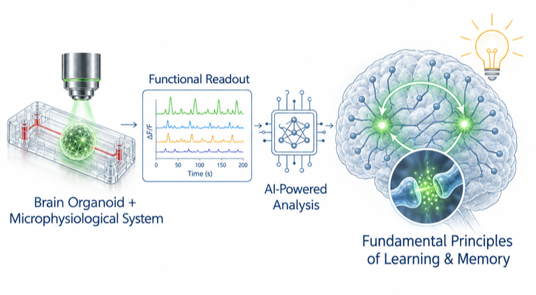
We address this challenge through engineering. By combining human stem cell biology, tissue engineering, microfluidics, and advanced multiphoton imaging, we build microphysiological systems that reconstruct key aspects of human brain development. These models let us observe how human tissues develop, communicate, and respond to perturbations in ways conventional models cannot. Our goal is not to miniaturize human tissue but to ask new questions about neuroscience for learning and memory and identify therapeutic strategies for neuromuscular and neurodevelopmental disorders.
Our favorite engineering and development tool
Microfludics & electrophysiological recording
Microfluidic devices with electrodes for recording neural activity.
See
Osaki et al., Nat. Comms, 2024
Osaki et al., Scientific Reports. 2018
Neuromuscular-on-a-chip
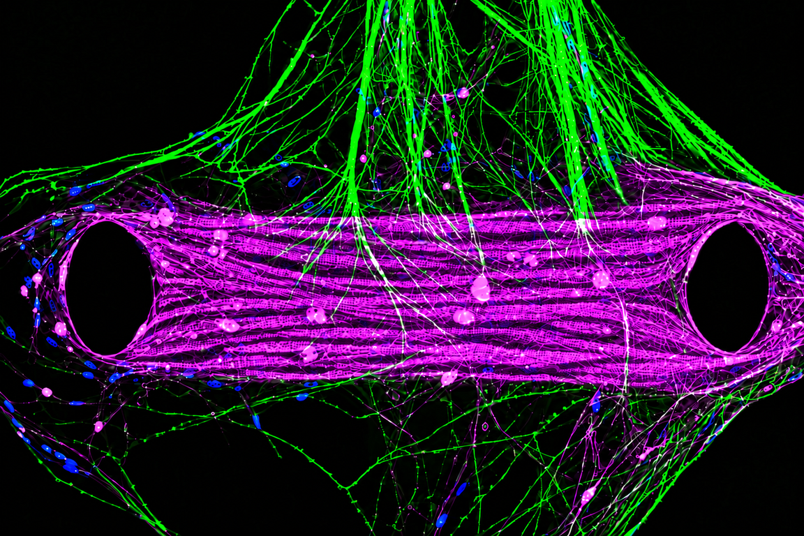
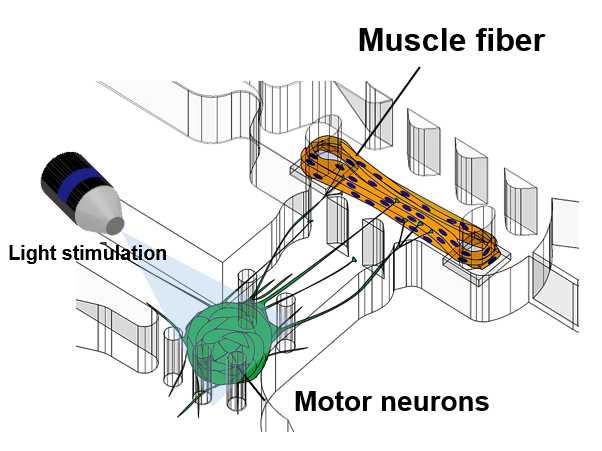
Motor neurons and muscle cells co-cultured on a chip to investigate the underlying mechanisms of ALS.
See
Osaki et al., Science Advances, 2018
Osaki et al., Nature Protocols, 2020
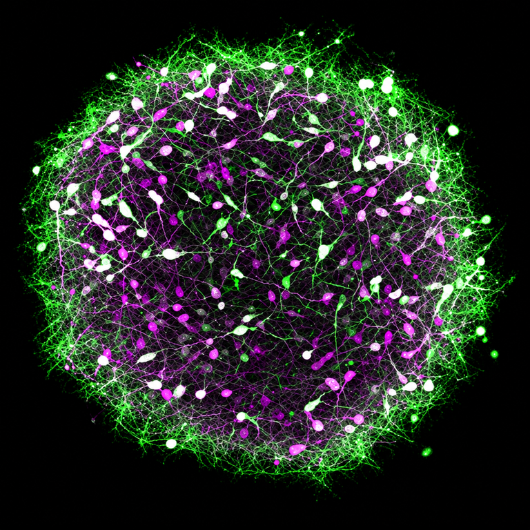
Genome editing in Stem cell and brain organoids
Engineering iPS cells and brain organoids to model human brain disease
See
Osaki et al., Nat. Comms, 2026
Osaki et al., Molecular Psychiatry. 2026
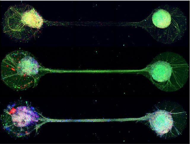
Advanced organoids-on-a-chip
Brain organoids connected on microfluidic chips to study neural circuits.
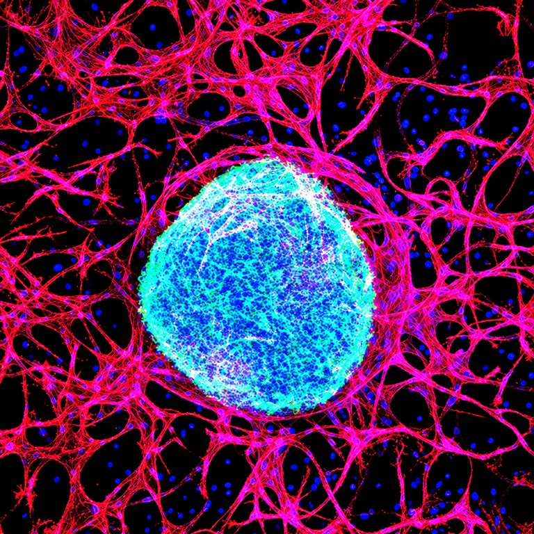
Neurovascular-on-a-chip
Models of neurons, glia, and blood vessels to study neurovascular interactions.
See
Osaki et al., Scientific Reports, 2018
Osaki et al., Molecular Psychiatry. 2026
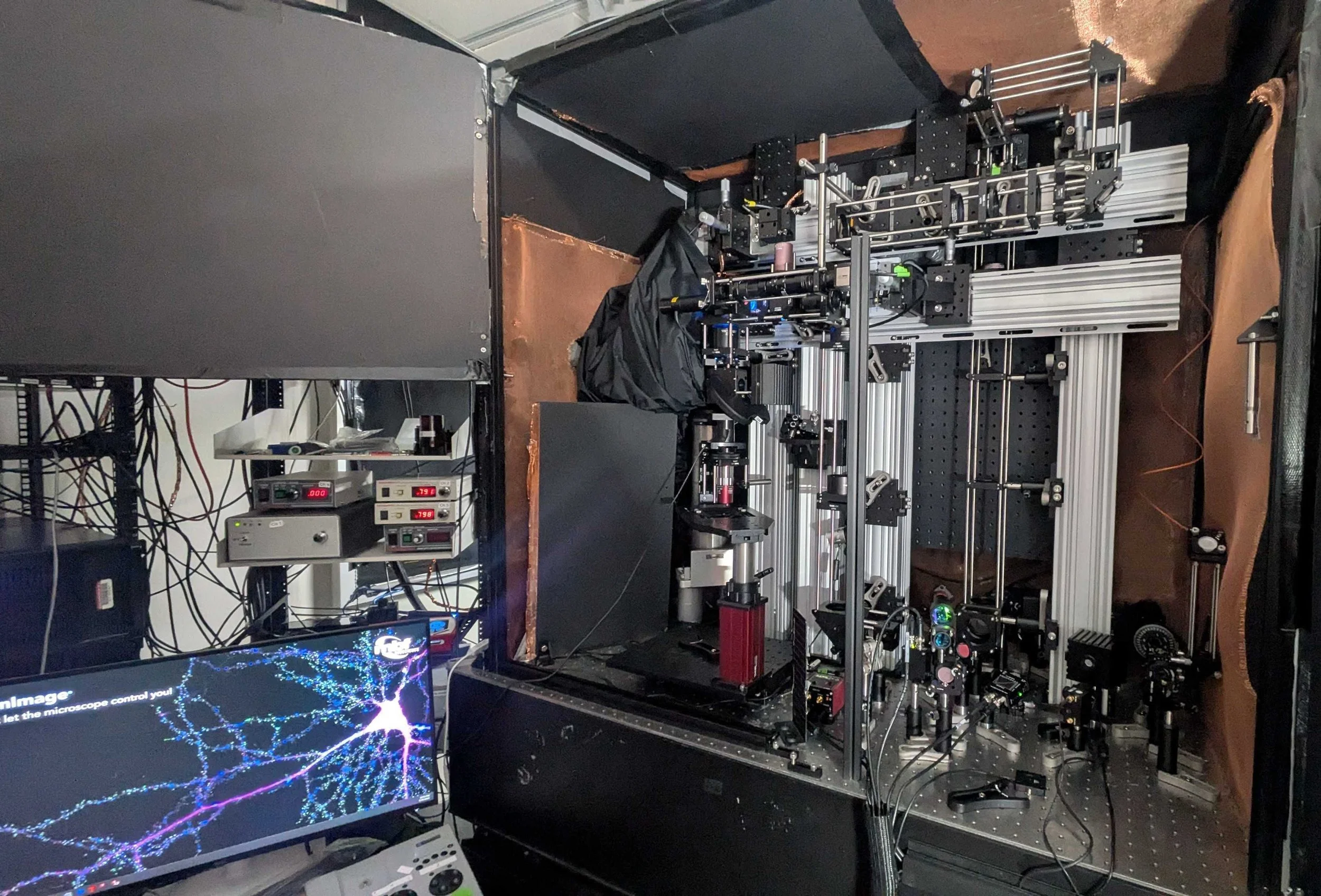
Advanced microscopy for
all optical electrophysiology
Multiphoton imaging with machine learning to decode neuronal activity.
See
Osaki et al., Light: Science& Applications, 2025
Our favorite questions
1.
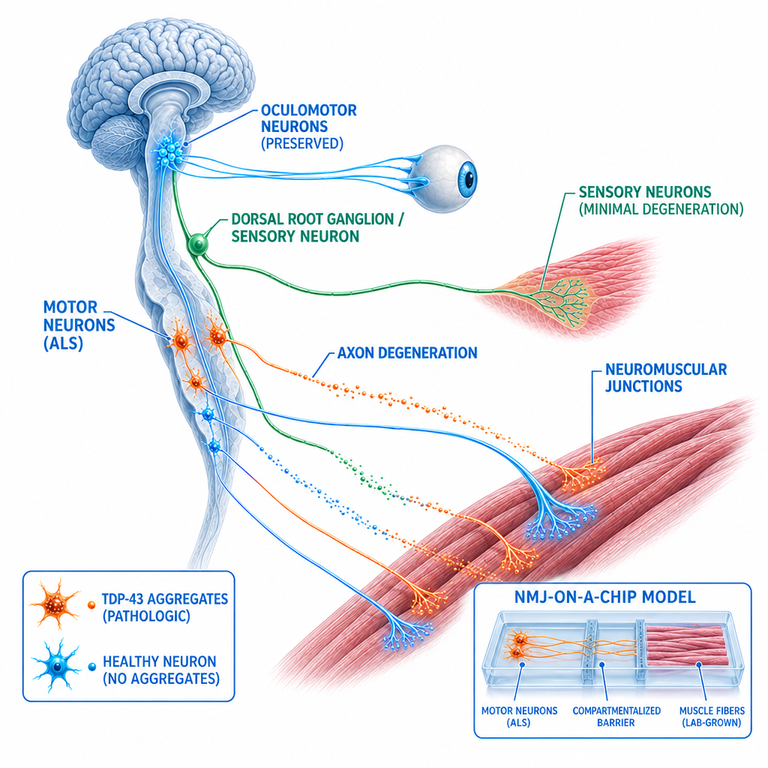
Motor Function and Its Disease-Related Dysregulation
How motor neuron degeneration were triggered in Amyotrophic lateral sclerosis?
- Why motor neuron is selectively died in ALS?
- What is the contribution of sensory neurons in ALS?
- How ocular motor neuron escape from neurodegeneration in ALS?
2.
Cortical Development and Plasticity
How long-range cortical input contribute to cortical plasticity & neurodevelopment?
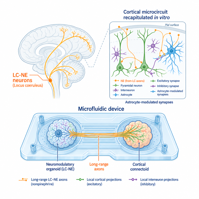
— How neuromodulatory projection from mid brain or hind brain contribute to modulate internal state in cortex.
— In neurodevelopmental disorder such as Rett syndrome and Autism spectrum disorder, how these projection contribute to corticogenesis and underlying the mechanism of parthenogenesis?
3.
Learning and Memory with AI and living system
How can we implement microphysiological system and AI virtual cell to understand fundamental principle of learning & memory?
- What is needed for in vitro engineered living system to be evaluated as model for clinical trial and drug screening